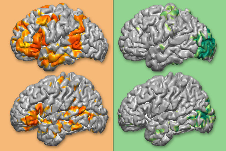

Timothy Mitchell, PhD (Instructor, Physics Division) provided data analysis assistance for a brain-mapping project that has recently been funded for 3.6 million dollars by the National Cancer Institute of the National Institutes of Health (NIH). Dr. Mitchell contributed to the study by mapping patients with large brain tumors using an algorithm created by graduate student Carl Hacker to create a cortical network map. “We were able to compare the motor and language areas of the brain as identified by Carl’s algorithm and compare it to electrocortical stimulation mapping that took place intraoperatively and found that there was good agreement between the two methods of network identification,” says Dr. Mitchell.
The multi-departmental study, led by Washington University faculty from Radiology and Neurosurgery, aims to develop software to aid in mapping patients’ brain function areas (ex. language or motor function) as well as anatomical structures, creating a better guide for neurosurgeons to use during operations.